All posts by Peter Brodersen

OCCM Seminar Series: Prof. Arthur Blackburn

‘Scanning Electron Diffraction at Sub-30 keV: Towards Democratization of High-Resolution Electron Microscopy’
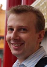
Prof. Arthur Blackburn
Dept. of Physics and Astronomy
University of Victoria

Time: Tuesday, August 6, 1-2pm
Location: Wallberg 215
Host: Jane Howe, jane.howe@utoronto.ca, 416.946.7221

In recent years, there is a strong interest in ptychography, which computationally constructions a model of the sample from series of diffraction data collected from electron microscopes [1]. We have demonstrated a resolution of 0.67 Å or better using ptychography with a 20 keV electron beam operating in a transmission mode using a SEM with a cold emission gun and immersion lens [2]. We achieved this through combination of: adding a simple diffraction projector lens to our SEM; using an un-coated hybrid direct electron detector [3]; and incorporating correction of diffraction projector distortions within a multi-slice ptychographic reconstruction process. Measurement and corrections of lens distortions is in general not trivial, so in our work we have simplified distortion evaluation using machine learning methods, as will be described. Also, using low energy scanning transmission electron diffraction in a SEM has provided us very valuable information for understanding the structure of novel polymer structures, as will be illustrated.
These advances serve to widen access to, and hence help democratize, information that is currently only available from high-end TEM. At low bean energies interesting possibilities are presented with computationally assisted SEM, particularly with imaging thin, low atomic-number based samples, which present greater information at lower energies. This may in the longer-term assist in the structural determination of proteins with masses below about 100 kDa.


References: 1MJ Peet, R Henderson, et al., Ultramicroscopy 203, 125-131 (2019); 2Submitted – under revision; 3G Tinti, H Marchetto, et al., J Synchrotron Radiation 24, 963-974 (2017).


Arthur Blackburn is the Hitachi High-Tech Canada Research Chair, Co-Director of the Advanced Microscopy Facility and Assistant Professor in the Department of Physics and Astronomy at the University of Victoria. Prior to joining the University of Victoria, he was a Senior Research Scientist in the Hitachi Cambridge Laboratory, embedded with the Cavendish Laboratory of the University of Cambridge. He progressed to this role after completing his PhD within the University of Cambridge, Department of Physics.

OCCAM Seminar Series: Dr. Jason Tam

‘Atomic Resolution Electron Microscopy Study of Grain Boundary Segregation Sequence in Yttria Stabalized Zirconia’

Dr. Jason Tam
Institute of Engineering Innovation
University of Tokyo

Time: Monday, August 12, 11am to noon
Location: Wallberg (WB) 407
Host: Jane Howe, jane.howe@utoronto.ca, 416.946.7221

Manipulating the structure and chemistry of grain boundaries and interphase interfaces in crystalline materials are crucial to obtain materials with desirable physical and functional properties. Although there are many experimental studies on grain boundary segregation in various alloys and ceramics, the study of the transformation from the initial solid solution structure at the atomic scale have not been explored. In this study, a novel bicrystal technique was developed to produce yttria stabilized zirconia (YSZ) bicrystal specimens without Y3+ segregation to the grain boundary. Scanning transmission electron microscopy (STEM) and energy dispersive X-ray spectroscopy (EDS) characterization confirmed that the specimen is indeed free of Y3+ segregation. To trigger grain boundary segregation, the specimen was annealed at various temperatures and the structure and chemistry were tracked by atomic resolution STEM imaging and EDS mapping. The fundamental understanding of the segregation sequence, as well as the conditions that can activate solute segregation are important for tailoring the properties and behaviour for the specific applications of YSZ, including solid electrolyte for solid oxide fuel cells (SOFCs) and cutting tools.

Jason Tam is currently a postdoctoral researcher at The University of Tokyo. He received his B.A.Sc. and Ph.D. from the Department of Materials Science and Engineering, University of Toronto. During his graduate study, he was also a visiting scholar at Hokkaido University and The University of Tokyo. Prior to his current position in Japan, he took on several roles at the University of Toronto as a postdoctoral researcher, undergraduate course instructor, and research scientist supporting the operations of the electron microscopy facility in OCCAM. His research interests include physical metallurgy, specifically interfaces of materials, electrochemical synthesis of nanostructured materials, and electron microscopy.

OCCAM Seminar Series: Prof. Yu-Tsun Shao

‘New Ways to See Real-Space Topological Textures and Their Order Parameters at the Atomic Scale’

Prof. Yu-Tsun Shao
Dept of Chemical Engineering & Materials Science
University of Southern California

Time: Thursday July 18, 2024, 11 am to noon
Location: Wallberg (WB) 215
Host: Jane Howe, jane.howe@utoronto.ca, 416.946.7221

Topological structures in ferroic materials can emerge as particle-like objects such as skyrmions and merons, with real-space swirling arrangements of the order parameter that not only have mathematical beauty but hold promise for potential applications in next generation nanodevices. As those ferroic textures are intrinsically nm-scale and dynamic, developing methods for visualizing and characterizing their detailed 3D structure is a critical step in understanding their properties and exploring possible phase transitions. I will show how the measurement of structural information such as polarization, strain, chirality, electric or magnetic fields was made possible by new imaging methods, i.e., four-dimensional scanning transmission electron microscopy (4D-STEM) diffraction imaging. I will report the observation of room temperature Néel-type skyrmion in a van der Waals ferromagnet accompanied by a change in crystallographic symmetry and chemical order. Second, I report the emergence of achiral polar meron lattice (topological charge of +1/2) from disordered but chiral skyrmion (topological charge of +1) phase transition driven by elastic boundary conditions. Further, using multislice electron ptychography, the 3D structural distortions of unknown polar textures in complex oxide heterostructures can be resolved at unprecedented resolution and precision.

Yu-Tsun Shao studies quantum materials by novel electron microscopy techniques, specifically 4D-STEM. He studies the (multi-)ferroic crystals with the aim to elucidate the microscopic origin of interactions among local polar/magnetic order, strain, and chiralities during topological phase transitions. Before joining USC, Yu-Tsun did postdoctoral work in Professor David Muller’s group at Cornell University and received his Ph.D. in Materials Science and Engineering at the University of Illinois at Urbana-Champaign in 2018, under the mentorship of Professor Jian-Min Zuo.

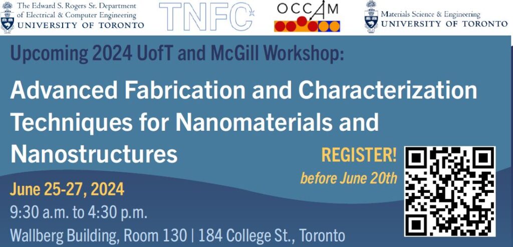
OCCAM and TNFC Co-host UofT/McGill Summer School on Advanced Fabrication and Characterization

The Summer School for Advanced Fabrication and Characterization Techniques is a three-day event from June 25 to 27, 2024, in Toronto Ontario, hosted by and the University of Toronto and McGill University through the Open Center for the Characterization of Advanced Materials (OCCAM) and the Toronto Nanofabrication Centre (TNFC). This summer school is the 2nd UofT-McGill Electron Microscopy Workshop.  Over the three days, participants can attend keynote talks from leading researchers showcasing the latest research trends on techniques and methodologies in electron microscopy, surface science, and semiconductor processing, including electron tomography, electron holography, volume electron microscopy, photolithography, and multiple high-end deposition and plasma etching systems.

Additional information, including a link to registration, can be found at The Summer School for Advanced Fabrication and Characterization Techniques .

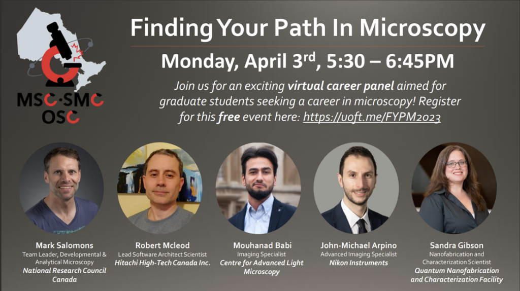
Microscopy Society of Canada Hosting Online Career Event, April 3/2023

The Microscopy Society of Canada Ontario Student Chapter is hosting an online career event Monday, April 3rd, 5:30 to 6:45 p.m. “Finding Your Path In Microscopy” is a virtual career panel that will feature microscopists currently working in industry, with the goal of highlighting career stories and opportunities for young microscopist trainees.

Panelists include:

  • Mark Salomons – National Research Council Canada
  • Robert Mcleod – Hitachi High-Tech Canada Inc.
  • Mouhanad Babi – Centre for Advanced Light Microscopy
  • John-Michael Arpino – Nikon Instruments
  • Sandra Gibson – Quantum Nanofabrication and Characterization Facility

If you are interested in attending, please register for free at the following link: https://uoft.me/FYPM2023.

Hope to see you there! 

OCCAM Joins Network of Micro-Nanotechnology Laboratories

CMC Microsystems is a not-for-profit organization accelerating research and innovation in advanced technologies.

OCCAM recently joined a network of Canadian open-access user facilities participating in CMC Microsystems’ micro-nanotechnologies (MNT) program. The MNT program provides financial assistance for custom microfabrication projects at open-access university facilities across Canada. Program eligibility extends to characterization work of the type in which OCCAM specializes. Details of the program are available here.

New SU7000 Installed in OCCAM

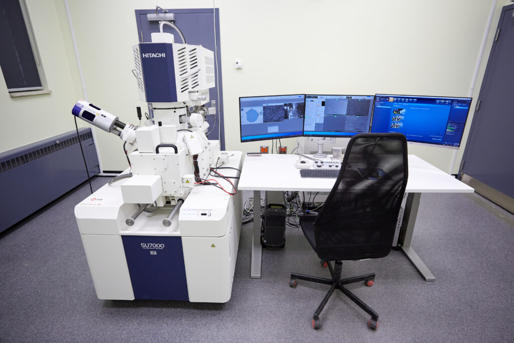
State-of-the-art SU7000 Ultra-high resolution scanning electron microscope installed in OCCAM’s Pratt laboratories.

A new Hitachi SU7000 FE-SEM instrument was recently installed in OCCAM. The ultra-high resolution variable pressure Schottky FE-SEM was purchased with a CFI-JELF awarded to Prof. J. Howe. The new instrument complements existing instrumentation within OCCAM and provides a key platform for development of new technologies and methodologies for in-situ characterization.

The SU7000’s optics and detector design, augmented by variable pressure capabilities, make it a highly capable and flexible tool for a wide variety of characterization modes and sample types. The instrument will eventually be equipped with energy dispersive x-ray analysis (EDX), electron backscatter diffraction (EBSD), and scanning transmission (STEM) capabilities.

The instrument will be available for applications work starting mid-November/2021. Hands-on user training will commence as soon as the public health situation allows.

OCCAM Adds New Hitachi HT7700 TEM/STEM

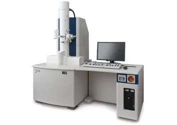
OCCAM has added a Hitachi HT7700 120keV TEM/STEM to its cadre of instrumentation.  The purchase was made possible through the generous support of three UofT engineering departments (Material Science & Engineering, Chemical Engineering & Applied Chemistry, Mechanical & Industrial Engineering).  The tool is installed in the Department of Chemistry’s Centre for Nanostructure Imaging (CNI).  The operation of the tool is a joint venture between OCCAM and CNI.

The new HT7700 complements existing tools, and meets the evolving demands of our extensive and diverse research community.  The HT7700 is suitable for 4-dimensional STEM (4D-STEM) research of soft materials, including bio-organisms (previously-fixed), organic macromolecules and polymers, thin films and other beam-sensitive matter.  The instrument will also be employed as a development platform for advancements in microscopy technology and applications.  The tool will not only support leading-edge materials and technical research programs, it will seed and support cross-faculty collaborative research endeavours.

For inquiries, please contact peter.brodersen@utoronto.ca.

To book the tool, please visit https://sites.chem.utoronto.ca/cni/content/booking

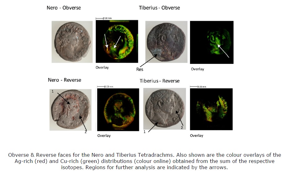
OCCAM staff apply xps/tof-sims to study of alexandrian tetradrachms

OCCAM research staff, in collaboration with Queen’s University Professor Cristiana Zaccagnino, have applied a combination of specialized material characterization techniques (electron microscopy and various surface spectroscopies) to study of Roman coins from the Diniacopoulos Collection of Central and Eastern Mediterranean Antiquities.

The work was recently published in Surface and Interface Analysis.

Volume/Number: Surf. Int. Anal., doi.org/10.1002/sia.6830

DOWNLOAD